HIE Brain Imaging
Brain Imaging For HIE Birth Injuries
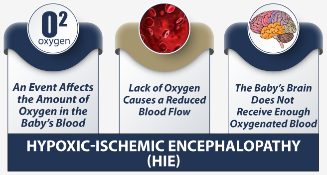
Hypoxic-ischemic encephalopathy (HIE) is a birth complication that can occur around the time of labor or delivery. HIE can lead to cerebral palsy or other severe neurologic defects in babies, and may cause the baby’s death.
HIE Neuroimaging
Imaging plays a critical role in the diagnosis and early medical intervention of HIE, thereby lowering the risks associated with HIE. Some of the key tools available for neuroimaging include magnetic resonance imaging (MRI), computed tomography (CT) and cranial ultrasound (CS).
The brain injury pattern in HIE depends on the duration and severity of hypoxia as well as the extent of the baby’s brain maturation. MRI, CT, and cranial US images can reveal the characteristic brain injury patterns associated with HIE to confirm the diagnosis and help predict neurological outcomes, such as cerebral palsy.
In a birth injury lawsuit involving an HIE brain injury, the MRI and other imaging is often important evidence that helps the birth injury lawyer “time” the injury and prove that HIE was avoidable had earlier intervention, like an emergency c-section, been performed.
How does an HIE injury continue to cause brain damage after birth?
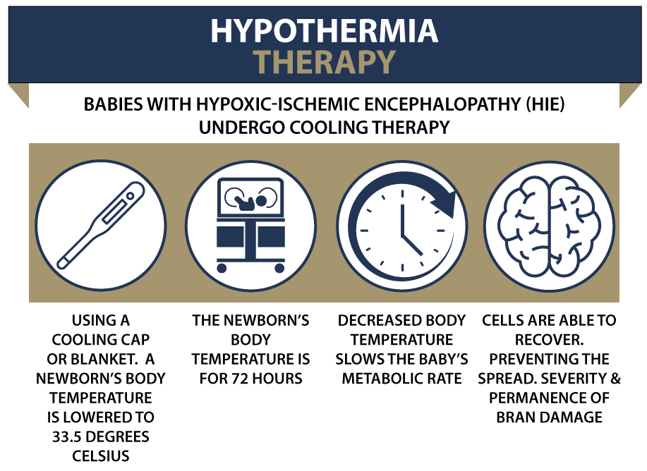
The management of a baby with suspected hypoxic-ischemic encephalopathy requires early medical intervention. This is because when a baby suffers hypoxic ischemic encephalopathy (HIE), the damage can progress even after birth if aggressive therapy (such as brain cooling or hypothermia therapy) is not initiated in order to minimize the brain damage.
Hypoxic-Ischemic Insult (HII)
Here is the general HIE timeline describing the major events that occur after the baby suffers a Hypoxic-Ischemic Insult (HII):
- Up to 12 hours after injury: The “acute” phase begins with significant involvement of the basal ganglia (structure near the brain center, responsible for motor control). It’s marked by focal cerebral lesions and focal cerebral infarction (aspects of cellular death).
- 12 to 24 hours after injury: Basal ganglia (BG) damage occurs in this phase, with nuclear pyknosis (degenerative changes) in the gray matter and coagulative necrosis (tissue death) in the white matter.
- 24 to 72 hours after injury: Significant signs of cellular swelling can be observed at this time due to brain edema (fluid buildup in the brain). The intra-cranial pressure (ICP) also increases, which can further injure the brain and spinal cord.
- 72+ hours after injury: This is the “sub-acute” phase of brain inflammation, marked by macrophage infiltration (to clear the dead brain cells and mineralization of the cellular residue). Selective nerve cell death may continue to occur in the basal ganglia, thalamus, and brain stem at this time.
MRI Tests can identify common brain injury patterns in HIE
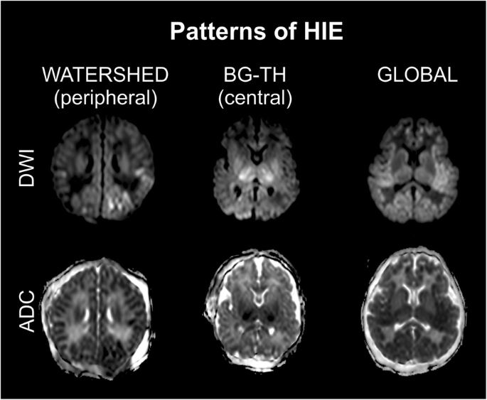
The location and extent of the brain injury in babies following birth asphyxia can vary according to the timing, duration, and severity of the hypoxic-ischemic insult. Based on human and animal studies, researchers have identified three major patterns of brain injury in babies with HIE:
Basal Ganglia and Thalamus (BGT) Injury Pattern
Severe fetal hypoxia or birth asphyxia particularly affects those areas of the baby’s brain that have the maximum metabolic demand for oxygen. These areas include the basal ganglia (responsible for motor function) and thalamus (responsible for sensory information relay, learning and memory).
Cerebral lesions in BGT are strongly associated with impairment of motor neurodevelopment and cerebral palsy. The more severe the BGT lesions, the more severe will be the motor impairment and the greater will be the likelihood of cerebral palsy.
Note: Lesions are an abnormal area of the tissue inside the body that may indicate underlying injury or disease.
White Matter/Watershed (WM/WS) Injury Pattern
When birth asphyxia is mild to moderate (or partially prolonged), the blood flow is able to reach the areas of the brain, which have the highest need for energy supplied through oxygen and glucose. But this happens at the cost of the watershed areas of the cerebral arteries – which means, the cerebral arteries become deficient in oxygen and glucose.
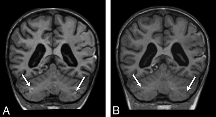
This causes an injury pattern is known as WM/WS or a parasagittal pattern of brain injury (distribution of injury along the brain’s vascular border zones). This damage is seen in MRI scans. In more severely injured babies, this brain injury pattern will also be seen in MRI scans in the subcortical white matter.
Initial neurological damage in babies is often limited in this type of injury, but neurological (“brain”) injury can worsen over time. Therefore, careful medical observation of the baby is important in these cases. An early follow-up between 12 to 18 months post-birth may indicate a normal outcome, but the baby is at risk of developing cognitive deficits with the passage of time. For this reason, long-term follow-up is recommended for a timely detection of disabilities and for making early interventions.
Near Total Injury Pattern
When the baby suffers from prolonged and very severe asphyxia (“profound”), it may result in near total brain injury, involving both the BGT (basal ganglia and thalamus) and white matter.
The incidence of this brain injury pattern is less frequently reported than BGT and WM/WS injury patterns simply because the baby may be too sick to be scanned with MRI after birth, or may die before the imaging tests can be performed.
The conventional MRI scans and even more advanced diffusion weighted imaging (DWI) scans may underestimate the extent of brain injury in these cases. Researchers increasingly point to a superior role of some of the latest MRI technologies, such as diffusion tensor imaging (DTI) to properly detect the brain injury.
HIE Diagnosis: The Role of Imaging
Neuroimaging tests, such as magnetic resonance imaging (MRI), computed tomography (CT) and ultrasound (US) can help in identifying and determining the precise location, severity, and extent of the brain injury in babies with HIE.
Advanced imaging tests, such as magnetic resonance spectroscopy (MRS), diffusion weighted imaging (DWI) and diffusion tensor imaging (DTI) are more sensitive to detecting brain injury and can play a key role in the timely diagnosis and early medical intervention to improve outcomes of babies suffering an HIE birth complication.
Magnetic Resonance Imaging (MRI)
Conventional MRI
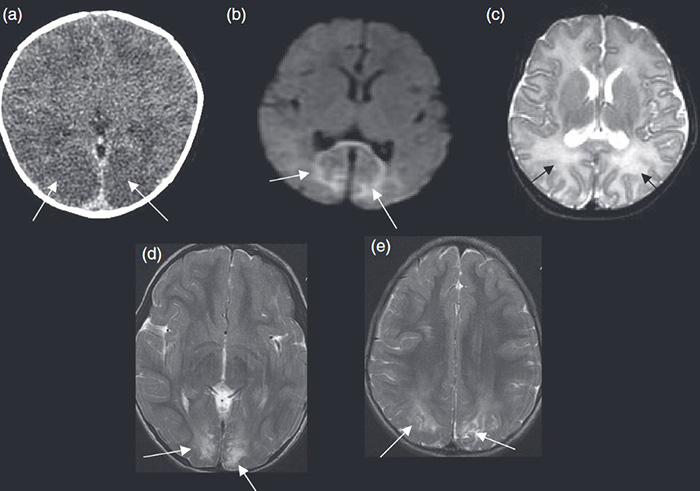
In the screening and evaluation of suspected HIE in babies, MRI is considered the gold standard because of its highly specific and sensitive imaging results. The MRI sequences commonly used in neonatal brain imaging are T1-weighted and T2-weighted scans, which are useful in evaluating the brain injuries in babies with fetal hypoxia and suspected HIE.
Note: Signal intensity of the tissue in an MRI scan refers to the “brightness” of the tissue revealed in the scan. The relative brightness (signal intensity) of the brain tissue indicates its condition or extent of damage to the medical observer.
Decreased T1 signal intensity indicates brain edema (as noticed in watershed damage in the white matter), while increased T2 signal intensity indicates a more severe (“profound”) neurological damage in the basal ganglia and thalamus (BGT).
HIE injuries will typically show on MRI:
- a. Increased basal ganglia signal intensity (T2)
- b. Increased thalamus signal intensity (T2)
- c. Decreased or non-existent signal intensity in the white matter (T1)
- d,e. Restricted water diffusion (when DWI technique is used for diffusion-weighted images)
When a baby has suffered an HIE birth injury, the abnormalities revealed in T1 and T2 MRI results are usually subtle in the first 72 hours after birth, and thereafter start becoming more apparent during the rest of the first week.
In other words, these injuries become more apparent and noticeable on an MRI 2-3 days after the HIE injury has occurred. This is important in allowing a neuro-radiologist to “time” the HIE birth injury.
T1 and T2 images in an MRI scan together reveal the lesions (abnormal tissue) associated with HIE (which is not visible through ultrasound). T1 images show the fat and soft-tissue anatomy (to determine the fat-containing mass).
On the other hand, T2 images reveal fluid anatomy and brain tissue abnormalities, such as brain trauma and inflammation. In practice, both T1 and T2 provide complementary information, and are reviewed together to characterize brain tissue abnormalities after an HIE injury.
Ultrasound and CT
In a suspected case of neonatal hypoxic-ischemic encephalopathy, the medical providers may use cranial ultrasound as the initial investigation technique during the first 24 hours (when an MRI may produce false-negative results—meaning that MRI does not reveal an HIE injury when one in fact exists).
A cranial ultrasound can successfully detect cystic PVL (brain tissue death around the ventricles), hydrocephalus (fluid buildup in the ventricles) and intracranial hemorrhage (brain bleeds).
On the other hand, ultrasound is not very accurate in detecting of cortical brain damage (injury to the cerebral cortex). The inter-observer variability in ultrasound exams is relatively high.
This means, there are differences in the ultrasound measurements between two different medical observers. In comparison, computed tomography (CT) has a higher sensitivity (sharpness or accuracy) than ultrasound for detecting cortical injury and a relatively low inter-observer variation.
However, CT has a lower sensitivity and specificity than MRI to detect HIE in babies. Nevertheless, a CT scan is useful in identifying intracranial bleeds in very sick babies without involving sedation. (In case of MRI, the newborn will have to be sedated.) The downside of CT, compared to ultrasound, is that CT involves significant radiation exposure for the newborn.
Magnetic Resonance Spectroscopy (MRS)
Nuclear magnetic resonance spectroscopy (MRS) is a more advanced technique than conventional MRI that can be used to identify and understand brain injury patterns that develop after hypoxic-ischemic encephalopathy.
In the deep gray matter of the brain, MRS measures the concentration levels of cerebral lactate and N-acetylspartate (NAA). Both of these measurements are important indicators of brain injury/damage.
When the baby’s brain is deprived of oxygen, it triggers chemical changes in the brain tissue leading to accumulation of cerebral lactate in the brain cells. Researchers have determined that distinctly elevated levels of lactate in the baby’s brain tissue are a key characteristic of birth asphyxia and an ischemic event.
Similarly, researchers have also established that low levels of NAA are indicated in a hypoxic/ischemic event in babies. With magnetic resonance spectroscopy (MRS), the concentration levels of lactate and NAA can be measured. Therefore, MRS has a good prognostic and predictive value in case of babies with suspected HIE.
MRI and MRS are different but complementary techniques, and can be used together to acquire a better understanding of the brain injury and the prognosis.
Diffusion Weighted Imaging (DWI)
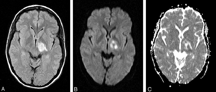
In a healthy brain, neurons absorb and release water molecules as they transmit messages throughout the brain. This process is known as water diffusion in the brain. When a hypoxic-ischemic insult (brain injury) occurs in the baby with HIE, this water diffusion becomes restricted.
This means, water accumulates in certain areas of the brain. This is the hallmark feature of HIE, which usually develops within one hour of the insult.
Diffusion weighted imaging (DWI) can show the restricted water diffusion (water accumulation) in the baby’s brain tissue. This lowering of the diffusion (measured as “apparent diffusion coefficient” or ADC) is a key determinant of the acute ischemic event (HIE) occurring in the baby.
The immediate reduction in ADC indicates brain swelling (cytotoxic edema) which usually takes places within minutes of the ischemic event.
DWI can show the areas of the baby’s brain with reduced ADC, which represents an irreversibly damaged brain tissue. Low ADC values with DWI (indicating brain injury) can be noticed at a much earlier stage when the T1 and T2 weighted sequences from a conventional MRI are still not visible.
Brain abnormalities noticed in DWI scans usually peak (meaning are most visible) within three to five days following the HIE birth injury and usual normalize (disappear) in about 11 to 12 days for babies who receive therapeutic hypothermia.
Diffusion Tensor Imaging (DTI)
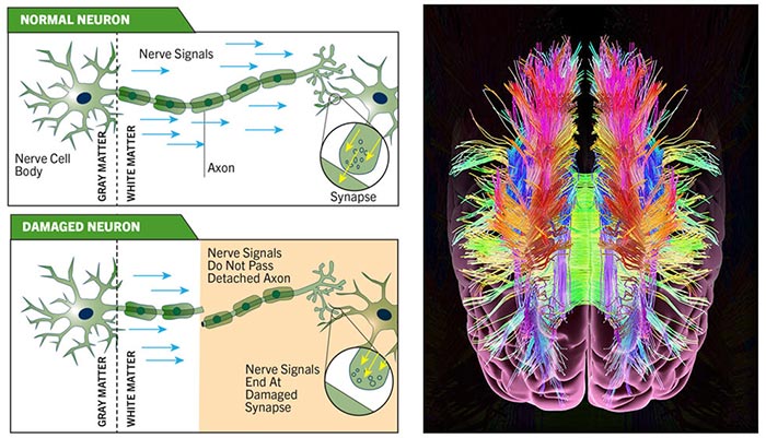
Water molecules in the brain do not diffuse uniformly through all structures of the brain. In case of white matter (present in the subcortical or deeper tissues of the brain), water diffusion occurs parallel to the white matter fibers.
During a hypoxic-ischemic event, the water diffusion not only gets restricted, but also changes its direction.
While Diffusion Weighted Imaging (DWI) can only measure the restricted diffusion (water accumulation), Diffusion Tenson Imaging (DTI) goes a step further and also measures the changes to the direction of the diffusion.
The analysis of the water diffusion pattern (known as anisotropy) helps trained radiologists evaluate the extent of ischemic, inflammatory, and traumatic brain tissue damage (lesions).
DTI can detect changes to the cortical neuron connections (cortex damage after white matter injury) – even when the conventional MRI images show normal brain structure and function (normal signal intensities).
Can DTI Predict Cerebral Palsy in Babies with HIE?
Multiple research studies have documented the diagnostic accuracy of DTI in case of babies with suspected HIE.
One study of 10 babies with HIE conducted within two weeks after birth found that DTI-based evaluation showed consistently abnormal results even when the measurements with MRI were totally unremarkable.
Another study involving 20 babies with HIE found that fractional anisotropy (FA) values – measuring the water molecules movement with DTI – were abnormal, even when ADC values (measured with DWI) showed normal results.
Based on the results of these studies, researchers suggested an imaging roadmap for babies with HIE as follows:
- Use conventional MRI with DWI for the prediction and early detection of cerebral palsy in cases of severe fetal hypoxia.
- Where mild to moderate HIE is suspected, but the MRI scan appears normal, perform DTI with accurate measurement and interpretation of FA values to lower the possibility of missing out cases of HIE and for an early prediction of cerebral palsy.
Researchers concluded that using advanced MRI tests such as DTI to evaluate term babies with HIE is a more accurate technique to predict neurodevelopmental outcomes, primarily cerebral palsy.
MRI Scoring System to Quantify Brain Injury
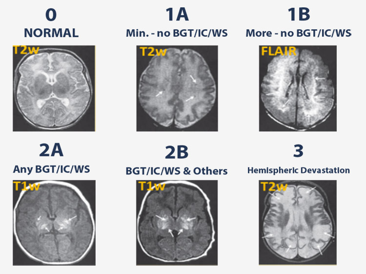
A number of MRI scoring systems have been proposed by researchers over the years to quantify the extent and severity of brain injury and predict the outcomes for babies with HIE. The Neonatal Research Network at the National Institute of Child Health and Human Development (NICHD) has developed a system that grades HIE brain injury in babies using six categories:
Note: Lesions are an abnormal area of the tissue inside the body that may indicate underlying injury or disease.
- Score 0 – Normal
- Score 1A – Cerebral lesions in sub-cortical areas
- Score 1B – More extensive lesions in sub-cortical areas
- Score 2A – Lesions in basal ganglia, thalamus and the internal capsule
- Score 2B – Lesions in 2A areas and cerebral areas
- Score 3 – Cerebral hemispheric devastation
How does the timing of a Brain MRI relate to Diagnosing and Timing of HIE?
For the correct interpretation of MRI results, the timing of MRI tests is critical. This is because brain injury patterns on different types of MRI tests will vary significantly during the first two weeks after the hypoxic-ischemic encephalopathy (HIE) birth complication because the actual brain damage may continue to evolve over this time period. The optimal timing of MRI remains a matter of controversy, but researchers agree that early MRI is preferred over delayed scanning.
Brain MRI
With the availability of advanced MRI techniques, such as DWI and DTI, it’s possible to detect brain abnormalities early before pseudo-normalization sets in. This means if brain imaging tests are delayed after an ischemic event, the MRI scans may show false-negative or pseudo results close to normal.
Therefore, with advanced DWI and DTI techniques, the MRI tests can be conducted quickly in a baby with suspected HIE, and there is a better possibility to prognosticate and predict neurodevelopmental outcomes, such as cerebral palsy.
According to researchers, MRI may also be performed during the application of therapeutic hypothermia without adversely affecting the therapy’s efficacy.
When there is a high suspicion of brain injury prior to birth, it is a key clinical indicator to perform MRI during the brain cooling process because this would allow for adjustment of the therapy for better outcomes.
It’s noteworthy that during the first 24 hours after the HIE birth injury, the MRI may provide false-negative results—meaning that even though an HIE injury has occurred it is not visible on the MRI at this time.
At this time, cranial ultrasound performed by trained medical professionals should be the first neuroimaging modality of choice to assess the presence of brain damage.
Brain CT
Computed Tomography (CT) should only be used in emergency when cranial ultrasound is not available because of the radiation-related future malignancy risks and the risk of damage to the baby’s intellectual development.
The Newborn Brain Society Guidelines and Publications Committee recommends that an MRI for babies with HIE should be performed immediately after therapeutic hypothermia (or two to five days after birth) to confirm the HIE diagnosis and predict outcome.
A second MRI (repeat scanning) is generally not required, but should considered at 10 to 14 days after birth if the medical providers find a difference between the early MRI results and the baby’s clinical condition, or if the first MRI led to ambiguous results. It is well-known that some baby’s MRI results may be negative after the completion of cooling treatment even though they have suffered from an HIE birth injury.
Clinical trials in recent years have shown that selective brain cooling in the form of therapeutic hypothermia helps in improving the neurological outcomes in babies with HIE.
Therefore, timely diagnosis and early medical intervention is the prime objective in the management of babies with suspected hypoxic-ischemic encephalopathy.
Is Your Child’s Birth Injury the Result of Medical Malpractice?
Parents whose children suffer from birth injuries or birth complications want and deserve answers as to cause of their child’s injury and whether mistakes by the doctors and nurses contributed to the injury.
- Were there signs of a birth injury during the pregnancy, labor, and delivery process, or presence of risk factors, which were either not recognized or properly treated?
- Did the medical team fail to order a series of tests to diagnose a suspected birth complication in a timely manner?
- Was the decision to perform a cesarean delivery delayed?
- During the labor and delivery, were there clear indications that their baby was suffering from fetal distress, but appropriate actions were not taken by the obstetrician or nurses?
- Did the neonatal resuscitation team delay in providing important breathing support after birth?
- Were serious neonatal conditions like hypoglycemia or jaundice missed or treated incorrectly?
- Should brain cooling (also called “hypothermia therapy”) have been offered to your baby, but the doctors and nurses failed to perform the appropriate tests or ignored the results of the tests?
Experienced Birth Injury Lawyers
The experienced birth injury lawyers at Miller Weisbrod Olesky, who have been through the legal battlefields before, will help you determine if mistakes of the medical providers caused a birth injury to your child, including Hypoxic-Ischemic Encephalopathy (HIE) or cerebral palsy.
Our dedicated birth injury attorneys have represented families all over the United States in their time of need after a birth injury. We use our experience and expertise to obtain you and your child a medical malpractice settlement that will help provide specialized medical therapy in order to maximize the quality of life and independence of your child throughout their life.
Sometimes families are reluctant to contact a medical malpractice lawyer. It’s also not uncommon for parents to feel overwhelmed by the responsibilities they encounter in caring for their injured child and worried that they will not be able to help out in a lawsuit involving their child’s birth injury.
Our birth injury attorneys and nursing staff will address these hesitations and concerns, so you can focus on your child and maximizing their care.
Registered Nurses and Nurse-Attorneys Are a Vital Part of Our Birth Injury Team…and Yours

Most birth injury law firms will employ one or two nurses to assist the review of cases and medical research. But Miller Weisbrod Olesky offers an unmatched number of nurses and nurse-attorney employees support to both the birth injury attorneys and our clients.
Our team of registered nursing staff and nurse-attorneys bring a deep level of medical and personal insight to every client’s case. Our nursing team includes both an experienced labor and delivery nurse as well as an ICU nurse. Working closely with the rest of the team, they investigate the reasons behind a birth injury and how medical professionals breached their standard of care.
Why Should You Talk with the Knowledgeable Attorneys at Miller Weisbrod Olesky?

The only way to find out if you have a birth injury case is to talk to a lawyer experienced in birth injury lawsuits. It’s not uncommon that a birth related complication results in a preventable birth injury, including cerebral palsy, but it takes a detailed expert review by a birth injury attorney of the medical records from your child’s birth to determine if the birth injury was the result of medical malpractice.
At Miller Weisbrod Olesky, a team of committed lawyers, nurses and paralegals uses our detailed medical negligence case review process to assess your child’s potential birth injury case. We start by learning more about you and your child and the status of meeting/missing developmental milestones. Then we gather medical records to determine what happened before, during, and after your delivery. We call in skilled medical experts who review your records and let us know if they think medical errors could have caused your child’s injuries.
If we feel medical negligence caused or contributed to your child’s injuries, we meet with you to discuss how you can receive compensation from the medical professionals who made the errors. Our birth injury attorneys have recovered millions of dollars in settlements for families of children that have suffered a birth injury.
At no point in our legal intake process will we ask you to pay anything. The medical review of your case and the consultation are free. We only receive payment when you do.
Contact Our Birth Injury Lawyers
National Birth Injury Law
Our National Birth Injury Attorneys, nurses, and support staff understand that parents of children with birth injuries feel overwhelmed. So, every client has the attention and support of a team of trained, compassionate professionals. But we don’t just offer compassion.
We offer a process to help you discover whether your child’s birth injury, HIE, cerebral palsy or brain injury at birth was caused by medical malpractice.
Call our offices today at (888) 987-0005 for experienced assistance in a free consultation.
Quick Links
Testimonials
- Lyric C. I feel like our voice was heard in a sense of what can possibly go wrong in a delivery and finding us answers. I feel with our settlement, we are now in a comfortable position to provide for our son.
- Lyssa L. They are not just people that say “hey let's get you money and let's go” The law firm was very thorough with us. It was awesome. I don't want to cry, because I think about and it's amazing that they were able to help me and that we were able to help my son and get the story out there.
- Jay C. Throughout the process, one thing was clear to us, the ultimate interest of our child was the utmost concern of Max and his team and as parents navigating a situation like that, that was refreshing to know we had them firmly on our side. I highly recommend them.
Popular Cities
- ★ Dallas Birth Injury
- ★ Houston Birth Injury
- ★ Atlanta Birth Injury
- ★ Chicago Birth Injury
- ★ Philadelphia Birth Injury
Locations
★ Dallas
11551 Forest Central Drive, Ste. 300
Dallas, TX 75243
★ Houston
12929 Gulf Freeway, Ste. 111
Houston, TX 77034